Anatomy Label Major Arteries And Veins - Detailed Labeled Anatomy Human Body | jpg: labeled heart ... : Jul 29, 2020 · there are three major types of blood vessels:
byAdmin•
0
Anatomy Label Major Arteries And Veins - Detailed Labeled Anatomy Human Body | jpg: labeled heart ... : Jul 29, 2020 · there are three major types of blood vessels:. Descending aorta coronary arteries pulmonary trunk pulmonary arteries jugular vein great cardiac vein right atrium left ventricle superior vena cava carotid artery Can you consciously control your breathing? Describe the basic process of hematopoiesis, where it occurs, and the significance of the pluripotent stem cell (hemocytoblast) in the process. Which type of vessels, arteries or veins, has more muscle fibers? This muscle is commonly confused as a rotator cuff muscle, but it is not because it does not attach to the capsule of the shoulder joint , unlike the teres minor muscle for.
Correctly label the following features of the aorta and its major branches. Which type of vessels, arteries or veins, has more muscle fibers? Blood vessels are often named after either the region of the body through which they carry blood or for nearby structures. Descending aorta coronary arteries pulmonary trunk pulmonary arteries jugular vein great cardiac vein right atrium left ventricle superior vena cava carotid artery In this animated and interactive object, learners identify the valves and chambers of the heart.
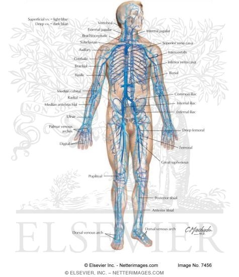
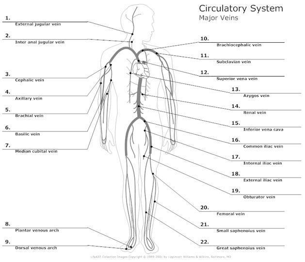
Major Veins of the Cardiovascular System from www.netterimages.com Jul 29, 2020 · there are three major types of blood vessels: Correctly label the following major systemic veins. The teres major muscle (from latin teres, meaning rounded) is positioned above the latissimus dorsi muscle and assists in the extension and medial rotation of the humerus. Correctly label the following features of the aorta and its major branches. The veins include the upper and lower vena cava system as well as the portal system. This muscle is commonly confused as a rotator cuff muscle, but it is not because it does not attach to the capsule of the shoulder joint , unlike the teres minor muscle for. One of its branches, the subclavian. Drag each label to the appropriate position to identify whether the structure contains oxygenated or deoxygenated blood.
This muscle is commonly confused as a rotator cuff muscle, but it is not because it does not attach to the capsule of the shoulder joint , unlike the teres minor muscle for.
For example, the brachiocephalic artery carries blood into the brachial (arm) and cephalic (head) regions. The major veins drain blood from the same organs and limbs that the major arteries supply. This muscle is commonly confused as a rotator cuff muscle, but it is not because it does not attach to the capsule of the shoulder joint , unlike the teres minor muscle for. Correctly label the following features of the aorta and its major branches. The veins include the upper and lower vena cava system as well as the portal system. Aug 07, 2020 · the arteries were superficially labeled, primarily as they form basic anatomical landmarks. In general, we have no conscious control over smooth muscle or cardiac muscle function, whereas we can consciously control to some extent all skeletal muscles. Describe the basic process of hematopoiesis, where it occurs, and the significance of the pluripotent stem cell (hemocytoblast) in the process. The anatomy of the heart by wendy dusek. Descending aorta coronary arteries pulmonary trunk pulmonary arteries jugular vein great cardiac vein right atrium left ventricle superior vena cava carotid artery Correctly label the following major systemic veins. Veins are blood vessels that bring blood high in carbon dioxide back to the heart. Correctly label the following major systemic arteries.
In general, we have no conscious control over smooth muscle or cardiac muscle function, whereas we can consciously control to some extent all skeletal muscles. The teres major muscle (from latin teres, meaning rounded) is positioned above the latissimus dorsi muscle and assists in the extension and medial rotation of the humerus. This muscle is commonly confused as a rotator cuff muscle, but it is not because it does not attach to the capsule of the shoulder joint , unlike the teres minor muscle for. Aug 07, 2020 · the arteries were superficially labeled, primarily as they form basic anatomical landmarks. Correctly label the following major systemic veins.
Circulatory System Diagram - Types of Circulatory System ... from wcs.smartdraw.com The major veins drain blood from the same organs and limbs that the major arteries supply. Can you consciously control your breathing? The teres major muscle (from latin teres, meaning rounded) is positioned above the latissimus dorsi muscle and assists in the extension and medial rotation of the humerus. Blood vessels are often named after either the region of the body through which they carry blood or for nearby structures. In this animated and interactive object, learners identify the valves and chambers of the heart. These general diagrams show the digestive system, with the major human anatomical structures labeled (mouth, tongue, oral cavity, teeth, buccal glands, throat, pharynx, oesophagus, stomach, small intestine, large intestine, liver, gall bladder and pancreas). Describe the anatomy of the aorta and its major branches and relate it with their functions. Correctly label the following major systemic arteries.
What is the functional significance of this?
Describe the basic process of hematopoiesis, where it occurs, and the significance of the pluripotent stem cell (hemocytoblast) in the process. Describe the features of blood that give it the characteristics of a connective tissue. Veins are blood vessels that bring blood high in carbon dioxide back to the heart. One of its branches, the subclavian. In this animated and interactive object, learners identify the valves and chambers of the heart. This muscle is commonly confused as a rotator cuff muscle, but it is not because it does not attach to the capsule of the shoulder joint , unlike the teres minor muscle for. These general diagrams show the digestive system, with the major human anatomical structures labeled (mouth, tongue, oral cavity, teeth, buccal glands, throat, pharynx, oesophagus, stomach, small intestine, large intestine, liver, gall bladder and pancreas). Jul 29, 2020 · there are three major types of blood vessels: Describe the anatomy of the aorta and its major branches and relate it with their functions. The veins include the upper and lower vena cava system as well as the portal system. Drag each label to the appropriate position to identify whether the structure contains oxygenated or deoxygenated blood. For example, the brachiocephalic artery carries blood into the brachial (arm) and cephalic (head) regions. Correctly label the following major systemic arteries.
This muscle is commonly confused as a rotator cuff muscle, but it is not because it does not attach to the capsule of the shoulder joint , unlike the teres minor muscle for. The major veins drain blood from the same organs and limbs that the major arteries supply. In general, we have no conscious control over smooth muscle or cardiac muscle function, whereas we can consciously control to some extent all skeletal muscles. In this animated and interactive object, learners identify the valves and chambers of the heart. The veins include the upper and lower vena cava system as well as the portal system.
Pin on Anatomy and Physiology Models from i.pinimg.com For example, the brachiocephalic artery carries blood into the brachial (arm) and cephalic (head) regions. These general diagrams show the digestive system, with the major human anatomical structures labeled (mouth, tongue, oral cavity, teeth, buccal glands, throat, pharynx, oesophagus, stomach, small intestine, large intestine, liver, gall bladder and pancreas). In general, we have no conscious control over smooth muscle or cardiac muscle function, whereas we can consciously control to some extent all skeletal muscles. Describe the basic process of hematopoiesis, where it occurs, and the significance of the pluripotent stem cell (hemocytoblast) in the process. One of its branches, the subclavian. Which type of vessels, arteries or veins, has more muscle fibers? What is the functional significance of this? The veins include the upper and lower vena cava system as well as the portal system.
What does this tell you
Veins are blood vessels that bring blood high in carbon dioxide back to the heart. Correctly label the following features of the aorta and its major branches. Describe the anatomy of the aorta and its major branches and relate it with their functions. Correctly label the following major systemic veins. The teres major muscle (from latin teres, meaning rounded) is positioned above the latissimus dorsi muscle and assists in the extension and medial rotation of the humerus. Aug 07, 2020 · the arteries were superficially labeled, primarily as they form basic anatomical landmarks. One of its branches, the subclavian. The veins include the upper and lower vena cava system as well as the portal system. Drag each label to the appropriate position to identify whether the structure contains oxygenated or deoxygenated blood. In general, we have no conscious control over smooth muscle or cardiac muscle function, whereas we can consciously control to some extent all skeletal muscles. Which type of vessels, arteries or veins, has more muscle fibers? Jul 29, 2020 · there are three major types of blood vessels: The major veins drain blood from the same organs and limbs that the major arteries supply.